
PrimeFlow™ RNA Assay reveals the dynamics of RNA and protein
expression within individual cells, facilitating unprecedented analysis
of their correlation as the cell changes over time or in response to stimulus.
This novel assay employs a proprietary fluorescent in situ hybridization
(FISH) technique enabling simultaneous detection of up to three RNA
transcripts in a single cell using a standard flow cytometer. RNA detection
may be teamed together with intracellular and cell surface antibody staining
to elevate the understanding of single cell dynamics to a new dimension.
Coupling RNA expression with protein detection on a flow cytometer generates
multiparametric data in heterogeneous cell populations and offers in-depth and
high content details at the single cell level. In contrast, microarrays and
sequencing can provide comprehensive gene expression data in bulk
sample preparations; however, such bulk sample preps can mask the
individual effects of different cellular subsets. Using the PrimeFlow™ RNA Assay,
specific cell populations may be analyzed for unique transcript expression levels
or cell subsets evaluated over time to determine transcriptional regulation and
protein expression simultaneously. Such unique and valuable insights are highly
applicable to answering previously unanswerable questions and have
broad implications for advancing research across multiple fields of biology.
Overview: PrimeFlow™ RNA Assay technology
Fluorescent in situ hybridization (FISH) is a powerful technique that allows
specific localization of ribonucleic acid targets in fixed cells. The basic premise
of the application relies on detecting nucleic acids through sequential hybridization
of nucleic acid probes, that provides to obtain gene expression information at a
single cell level. Current FISH techniques are generally limited by high background
and low sensitivity due to non-specific binding and inefficient signal amplification.
PrimeFlow™ RNA Assay incorporates a proprietary oligonucleotide probe set
design and branched DNA (bDNA) signal amplification technology to analyze RNA
transcripts by flow cytometry. bDNA technology provides a unique approach to RNA
detection and signal amplification by amplifying the reporter signal rather than the
target sequence (e.g. PCR) for consistent results, a common problem for PCR-based
assays.
In the PrimeFlow™ RNA Assay, target-specific probe sets contain 20-40 oligonucleotide
pairs that hybridize to the target RNA transcript. Signal amplification is achieved through
specific hybridization of adjacent oligonucleotide pairs to bDNA structures, formed by pre-
amplifiers, amplifiers and fluorochrome-conjugated label probes, resulting in excellent
specificity, low background and high signal-to-noise ratio.
PrimeFlow™ RNA Assay principle
The assay workflow contains several steps: surface antibody staining, fixation and
permeabilization, intracellular antibody staining, followed by target probe hybridization
with RNA-specific probe sets, signal amplification using bDNA constructs, and
detection by flow cytometry. For simplicity, detection of only two RNA targets are shown
in orange and yellow (in the figure above) with only three of the 20-40 oligonucleotide
target probe pairs per target RNA.
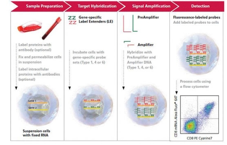
Sample preparation: Antibody staining, fixation and permeabilization
Single-cell suspensions can be stained for cell surface markers and fixable viability dyes,
as before the cells are fixed and permeabilized. Subsequently, the cells may be stained
with antibody directed to intracellular targets, such as transcription factors and cytokines.
After an additional fixation step, the cells are ready to proceed through the hybridization
and signal amplification steps.
Target hybridization
A target-specific Probe Set contains 20-40 oligonucleotide pairs that hybridize to specific
regions across the target RNA sequence. Subsequent signal amplification requires that
each half of a given oligonucleotide pair binds to the target RNA in adjacent positions.
Three types of probe sets are currently available to allow detection of RNA labeled with
Alexa Fluor® 647 (Type 1 Probe Sets), Alexa Fluor® 488 (Type 4 Probe Sets), or Alexa
Fluor® 750 (Type 6 Probe Sets). When detecting more than one RNA target in a single
sample, each Probe Set must be a unique type to differentiate its signal from the others.
Probe Set Type |
Fluorochrome Label |
Excitation Wavelength(max) |
Emission Wavelength (max) |
Laser Excitation Wavelength |
Bandpass Filter Recommendation |
Type 1 |
Alexa Fluor® 647 |
647 nm |
668 nm |
632-640 nm |
660/20 |
Type 4 |
Alexa Fluor® 488 |
488 nm |
519 nm |
488 nm |
530/30 |
Type 6 |
Alexa Fluor® 750 |
749 nm |
775 nm |
632-640 nm |
780/60 |
Signal amplification
Signal amplification using bDNA technology is achieved through a series of sequential hybridization steps, that forms a "tree" like structure. Pre-amplifier molecules hybridize to their respective pair of bound oligonucleotide probes to form the “trunk” of the tree. Multiple amplifier molecules hybridize to their respective pre-amplifier to create the “branches.” Finally, multiple Label Probes hybridize to the Amplifiers and form the "leaves" of the "tree." A fully assembled signal amplification tree contains 400 label probe binding sites. If all target-specific oligonucleotides in the probe set bind to the target RNA transcript, an 8,000 fold amplification can be achieved.
Fluorescence detection
Upon completion of the assay protocol, target RNA data is detected in cells by analyzing the sample on a standard flow cytometer equipped with a 488 nm & 633nm lasers and appropriate filter configurations, to capture the fluorescent signals.
FlowRNA Probe Sets
Choose from Thousands of Target-Specific Probe Sets
Selection of a Probe Set to detect RNA using the PrimeFlow™ RNA or QuantiGene® FlowRNA Assays is virtually unlimited. Each synthesized set of target probes typically spans 1000 bases (1 kb) of a target RNA. There are exceptions when the targeted RNA requires a smaller region or is expressed at low levels.
A Probe Set type is determined by its tag, a specific DNA sequence that generates a highly specific interaction with its own PreAmplifier, Amplifier and Label Probe. Three Probe Set types are currently available (Type 1, Type 4 and Type 6). When detecting more than one RNA target in a single cell, each Probe Set must be a unique type to differentiate its’ signal from the other Probe Sets. This is similar to cells labeled with multiple conjugated antibodies. Detection requires signals to be captured in individual channels on the flow cytometer using specific laser and filter set configurations (see table below).
Probe Set Type selection for Prime FlowRNA and QuantiGene FlowRNA assays should use the following criteria:
- Select Type 1 (Alexa Fluor® 647) when using one probe set in an assay to obtain the brightest signal. Select Type 6 (Alexa Fluor® 750) when adding a second probe set and finally Type 4 (Alexa Fluor® 488) is recommended for the third probe set.
- When multiplexing RNA targets, do not use Probe Sets of the same Type.
Currently, up to three compatible fluorochrome-labeled amplification “tree” structures allow simultaneous measurement of up to three different RNA targets. For each RNA target, a unique probe set type must be selected in order to detect it independently of the others.
FlowRNA Probe Set Type Specifications
Probe Set Type |
Fluorochrome Label |
Excitation Wavelength(max) |
Emission Wavelength (max) |
Laser Excitation Wavelength |
Bandpass Filter Recommendation |
Type 1 |
Alexa Fluor® 647 |
647 nm |
668 nm |
632-640 nm |
660/20 |
Type 4 |
Alexa Fluor® 488 |
488 nm |
519 nm |
488 nm |
530/30 |
Type 6 |
Alexa Fluor® 750 |
749 nm |
775 nm |
632-640 nm |
780/60 |
Probe set volume requirements can be determined by the number of assays required.
- 120 µL = ~ 24 assays
- 600 µL = ~ 100 assays
- Larger sizes can be made available.
Over 3000 sequences used in microarray or sequencing assays are currently in stock, and other RNA targets including most mRNA or noncoding RNA sequences can be designed and manufactured for use in the FlowRNA assay in five business days or less.